When They Cooperate, Proteins Become Red
Max Planck researchers develop another tool for making the interactions between proteins visible
A cell's life processes are based on the interplay of proteins, genes, and smaller molecules. Biologists call these biochemical interactions a "molecular network". Now, scientists from the Max Planck Institute for Plant Breeding Research in Cologne, Germany, along with colleagues from the University of Cologne, have observed - live and in colour - proteins completing tasks together. They separated a red fluorescent protein (RFP) into two parts which can attach to other proteins. If two of these other proteins interact, the two halves of the RFP also attach. The RFP lights up red, fifteen times brighter than before, because the biologists have biochemically optimised the original protein. This colouring helps scientists investigate the connections in the molecular network (Nature Methods, July 21, 2006).
, newly developed by scientists in Cologne, can indicate protein interactions in cells. For this reason, they split the RFP into two parts, which they attached to two green fluorescent proteins. The green proteins were interacting with each other, so the two halves of the RFP also came together - and the protein turned red.")
Genome research has allowed biologists to achieve a good overview of which genes and proteins individual organisms work with. But this list of inventories does not suffice to understand the processes in cells. Normally, many genes and proteins work together to achieve a certain task. Cell and molecular biologists now hope to determine which biomolecules cooperate when, and how.
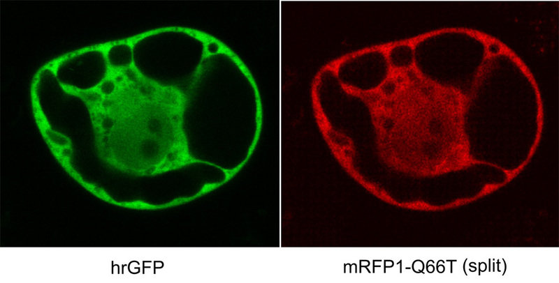
Scientists from the Max Planck Institute for Plant Breeding Research in Cologne and the University of Cologne have developed another tool for observing proteins as they cooperate. The scientists, led by Drs Guido Jach and Joachim Urig, have split an improved red fluorescent protein (RFP, fully denoted mRFP1-Q66T) into two parts which do not light up on their own. The two halves do not spontaneously find each other in a cell, but rather only when they are near to each other - that is when both are attached to a proteins that interact with each other. Then, the two RFP halves come together and light up red. The red fluorescence, which can be observed in a light microscope without destroying the cells, shows biologists that two prepared proteins are interacting with each other.
Biologists have been researching this process in cells for quite some time, with fluorescent proteins which cling to other proteins and light up when they are stimulated with the appropriate light. They can now combine the newly developed methods with systems that show the protein interactions with yellow, green or blue lights. They can follow, inside a cell, simultaneously, if and how various proteins come into contact with each other.